The human mouth, often taken for granted, is an intricate and dynamic gateway, serving as the initial point of contact for nourishment and a primary instrument for communication. It’s a complex anatomical region, a bustling hub of activity where several distinct structures work in concert to perform a multitude of vital functions. From the subtle movements that shape our words to the powerful actions involved in breaking down food, every part of the mouth plays a specific and indispensable role. Exploring these components reveals a marvel of biological engineering, perfectly adapted to its diverse responsibilities.
Outer Sentinels: The First Encounter
The structures forming the external boundary of the mouth are not merely passive barriers; they are active participants in its daily operations. These features are our first line of interaction with the world, both in terms of taking in sustenance and expressing ourselves.
The Lips: Expressive and Essential
Guarding the very entrance to our oral world are the lips (labia), remarkably mobile and sensitive structures. Composed of muscle, skin, and mucous membrane, they are far more than just gatekeepers. The lips play a crucial role in how we articulate sounds, enabling the formation of various consonants and vowels essential for speech. Their rich nerve supply makes them highly sensitive to touch and temperature, aiding in the assessment of food before it fully enters the mouth. Furthermore, the lips are instrumental in creating a seal for sucking, drinking, and holding food within the oral cavity during chewing. Their ability to convey a wide range of emotions, from a smile to a frown, underscores their importance in non-verbal communication. The distinct reddish hue of the lips, particularly in the area known as the vermilion border, is due to the presence of numerous blood vessels close to the thin surface epithelium.
The Cheeks: Walls of the Oral Chamber
Forming the lateral walls of the oral cavity are the cheeks (buccae). These fleshy structures are primarily composed of the buccinator muscles, along with subcutaneous fat and skin externally, and a mucous membrane internally. The cheeks work in close coordination with the tongue and lips to keep food positioned between the teeth during mastication, preventing it from spilling into the space between the teeth and the cheek lining. They also contribute to actions like sucking and blowing. The inner lining of the cheeks, the buccal mucosa, is kept moist by minor salivary glands, contributing to the overall lubrication of the mouth and aiding in the smooth passage of food.
Stepping Inside: The Oral Cavity Proper
Beyond the lips and cheeks lies the oral cavity proper, the main chamber of the mouth. This is where the majority of the mouth’s intricate machinery is housed, each component finely tuned for its specific tasks in digestion, speech, and even breathing to some extent.
The Dental Lineup: Our Tools for Mastication
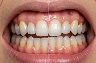
Central to the mouth’s function of processing food are the teeth (dentes). These hard, calcified structures are anchored in sockets within the alveolar processes of the maxilla (upper jaw) and mandible (lower jaw). Humans have two sets of teeth during their lifetime: the primary (deciduous or “baby”) teeth, and later, the permanent (adult) teeth. Each tooth consists of a crown, the visible part, and one or more roots embedded in the jawbone. The different shapes of teeth reflect their specialized roles in breaking down food.
Incisors: The Cutting Edge
Located at the front of the mouth, the incisors are typically sharp, chisel-shaped teeth. There are usually eight incisors in the adult dentition, four in the upper jaw and four in the lower. Their primary function is to bite into and cut food into smaller, more manageable pieces. Think of them as the initial slicers, preparing food for further processing by the teeth further back.
Canines: The Piercing Points
Positioned at the “corners” of the dental arches, next to the incisors, are the canines. Adults normally have four canines, two upper and two lower. These teeth are characterized by their pointed, conical shape, making them ideal for tearing and gripping food, particularly tougher items like meat. Their robust roots provide strong anchorage for these forceful actions.
Premolars and Molars: The Grinding Crew
Behind the canines are the premolars (or bicuspids) and molars. Premolars, typically eight in number, have broader, flatter surfaces with two cusps (points) and are used for crushing and grinding food. Further back still are the molars, the largest and strongest teeth, usually twelve in number (including wisdom teeth, which may or may not erupt). Molars have multiple cusps and extensive grinding surfaces, perfectly designed for the thorough pulverization of food before swallowing. This grinding action significantly increases the surface area of food, facilitating the subsequent stages of digestion.
The adult human typically possesses 32 permanent teeth, meticulously designed for different aspects of food processing. This complex dental toolkit, comprising incisors, canines, premolars, and molars, underscores the mouth’s primary role in initiating nutrition. Each tooth is anchored firmly within the jawbone, supported by the surrounding gums.
The Gums: Protective Foundations
Surrounding the necks of the teeth and covering the alveolar bone are the gums (gingivae). This specialized soft tissue is a dense, fibrous mucosa, firmly attached to the underlying bone. The gums provide a protective seal around the teeth, preventing food particles and microorganisms from reaching the sensitive tooth roots and supporting bone structures. The tissue is rich in blood vessels, giving it a characteristic pinkish appearance when in a state of normalcy. Proper interaction between the teeth and gums is essential for maintaining the stability and integrity of the entire dental apparatus.
The Tongue: A Muscular Marvel
Dominating the floor of the mouth is the tongue (lingua), a highly mobile and versatile muscular organ. It’s composed of a complex arrangement of intrinsic muscles (which alter its shape) and extrinsic muscles (which alter its position). The tongue is indispensable for numerous functions. It manipulates food during chewing, mixing it with saliva and positioning it between the teeth. It plays a crucial role in initiating swallowing by forming a bolus (a ball of chewed food) and pushing it towards the back of the mouth. Furthermore, the tongue is vital for speech, articulating a vast array of sounds by changing its shape and position against the teeth, palate, and other oral structures. Its surface is also equipped for another critical sense: taste.
A Landscape of Sensation: Papillae
The dorsal (upper) surface of the tongue is not smooth but covered with tiny projections called papillae. There are several types of papillae, including filiform, fungiform, circumvallate, and foliate papillae. Most of these papillae, particularly the fungiform, circumvallate, and foliate types, house taste buds. Taste buds are sensory organs that allow us to perceive the five basic tastes: sweet, sour, salty, bitter, and umami. The filiform papillae, which are the most numerous, do not contain taste buds but provide a rough texture to the tongue, aiding in gripping and manipulating food. This intricate surface helps in the mechanical processing of food and provides critical sensory information.
The Roof Over Our Mouths: The Palate
Forming the roof of the oral cavity and separating it from the nasal cavity above is the palate. It is divided into two distinct parts: the hard palate at the front and the soft palate at the back.
The Hard Palate: A Sturdy Platform
The anterior two-thirds of the palate is the hard palate. It is composed of bony plates from the maxilla and palatine bones, covered by a thick mucous membrane. This rigid structure provides a firm surface against which the tongue can press food during chewing and swallowing. It also features irregular ridges called rugae, which may assist the tongue in manipulating the food bolus.
The Soft Palate and the Uvula: Flexible Guardians
Posterior to the hard palate is the soft palate, a mobile, muscular flap of tissue that lacks a bony core. It is flexible and can be elevated or depressed by its associated muscles. During swallowing, the soft palate elevates to close off the nasopharynx (the upper part of the throat behind the nose), preventing food and liquids from entering the nasal cavity. Suspended from the midline of the posterior edge of the soft palate is a small, fleshy, conical projection called the uvula. The uvula also plays a role in sealing off the nasopharynx during swallowing and is involved in articulating certain sounds in speech. Its precise functions are still a subject of some study, but it contributes to guiding food downwards and plays a part in the gag reflex.
The Unseen Helpers: Salivary Glands
While not strictly part of the oral cavity’s lining, the salivary glands are crucial accessory organs that continuously produce and secrete saliva into the mouth. Saliva is a complex fluid essential for maintaining oral comfort and performing several key functions. It moistens and lubricates food, making it easier to chew and swallow. Saliva contains enzymes, such as amylase, which begin the chemical digestion of starches even before food leaves the mouth. It also helps to cleanse the mouth by washing away food debris and has properties that buffer acids and control microbial populations. There are numerous minor salivary glands scattered throughout the oral mucosa, but three pairs of major salivary glands produce the bulk of saliva.
Major Glandular Contributors
The three major pairs are the parotid glands, located near the ears, whose ducts open into the cheek opposite the upper molars; the submandibular glands, situated beneath the floor of the mouth, with ducts opening near the lingual frenulum under the tongue; and the sublingual glands, found under the tongue and anterior to the submandibular glands, which empty via multiple small ducts into the floor of the mouth. Together, these glands ensure a constant supply of saliva, adapting its flow and composition based on various stimuli, such as the sight, smell, or thought of food.
Supporting Structures: Floor and Connections
The floor of the mouth lies beneath the mobile part of the tongue and is formed by several muscles, primarily the mylohyoid muscles, which create a muscular diaphragm. This area houses the sublingual salivary glands and the ducts of the submandibular glands. It is covered by a thin mucous membrane. Connecting various mobile parts of the mouth to more fixed structures are folds of mucous membrane called frenula (singular: frenulum).
The Lingual Frenulum and Labial Frenula
The lingual frenulum is a prominent fold of tissue that anchors the underside of the tongue to the floor of the mouth. Its length and elasticity can affect tongue mobility. The labial frenula (superior and inferior) are folds that connect the inner surface of the upper and lower lips to the gums between the central incisors. Smaller buccal frenula can also connect the cheeks to the gums in the premolar regions. These tethers help to stabilize the lips and tongue while still allowing for their necessary range of motion.
The Vestibule: The Antechamber
An often-overlooked part of the mouth is the oral vestibule. This is the slit-like space situated externally to the teeth and gums, and internally to the lips and cheeks. Think of it as the antechamber of the oral cavity proper. When the mouth is closed and teeth are together, the vestibule is a closed-off space, communicating with the outside world only through the oral fissure (the opening between the lips). The ducts of the parotid salivary glands open into the vestibule. This space plays a role in holding food temporarily and allows for the movements of the lips and cheeks independent of the teeth and tongue to some extent.
Transition Zone: The Oropharynx
At the very back, the oral cavity transitions into the oropharynx, which is the part of the pharynx (throat) located behind the mouth. This junction is marked by structures like the palatoglossal arches (anterior pillars of the fauces) and the palatopharyngeal arches (posterior pillars of the fauces). Between these arches, on each side, are often located the palatine tonsils, which are part of the body’s lymphatic system. The oropharynx serves as a common passageway for both food heading to the esophagus and air heading to the larynx and lungs.
In essence, the human mouth is far more than a simple opening. It is a highly sophisticated and coordinated system of diverse parts, each contributing to fundamental life processes. From the initial bite to the articulation of complex thoughts, the mouth’s anatomy reflects a remarkable adaptation for survival, communication, and interaction with our environment. Understanding its various components allows for a deeper appreciation of this everyday marvel.