We often take them for granted, those thirty-two (or fewer, for some of us) champions of mastication nestled in our jaws. But human teeth are far more than just bite-sized tools for grinding down our food. They are intricate, living (in part!) structures, each a miniature marvel of biological engineering. Their design is a testament to efficiency and resilience, a complex interplay of hard and soft tissues working in concert. To truly appreciate them, we need to peel back the layers, metaphorically speaking, and explore the amazing architecture within.
The Outer Defenses: Enamel and Dentin
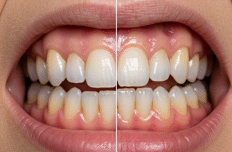
What we see when we smile is primarily the enamel, the tooth’s glossy, white outer covering. It’s not just for show; enamel is the hardest substance in the human body. Think of it as the tooth’s personal suit of armor. Its incredible strength comes from a densely packed matrix of mineral crystals, primarily hydroxyapatite. This mineral shield is tasked with withstanding the immense pressures of biting and chewing, as well as protecting the sensitive inner layers from the daily barrage of temperature changes and food acids. Interestingly, enamel contains no living cells, which means that once it’s significantly damaged or worn away, the body cannot regenerate it. This makes its preservation paramount.
Tooth enamel is indeed the hardest substance in the human body, surpassing even bone in its resilience. This remarkable strength is attributed to its highly organized, crystalline mineral structure. Despite its toughness, enamel is acellular, meaning it lacks living cells and thus cannot repair itself from substantial damage. Understanding this underscores the importance of protecting this vital outer layer.
Beneath this formidable enamel shell lies the dentin. If enamel is the armor, dentin is the sturdy framework that makes up the bulk of the tooth. It’s a bone-like material, but softer than enamel, and typically has a yellowish hue, which can become more apparent if the overlying enamel thins. Dentin is not a solid, inert mass; it’s permeated by thousands of microscopic channels called dentinal tubules. These tubules run from the enamel-dentin junction (or cementum-dentin junction in the root) towards the tooth’s core. Inside these tiny tunnels are fluid and extensions of cells that originate from the tooth’s pulp. This intricate network is why, if enamel is breached, you might experience sensitivity to hot, cold, or sweet stimuli – these sensations are transmitted through the tubules to the nerve center of the tooth.
The Living Core: The Pulp
At the very heart of every healthy tooth resides the pulp. This is the tooth’s living core, a soft tissue chamber filled with an intricate network of blood vessels, nerves, and connective tissue. The pulp chamber is the space within the crown (the visible part of the tooth), and it extends down through channels in the roots called root canals. The primary role of the pulp is formative; it’s responsible for producing the dentin as the tooth develops. Once the tooth is mature, the pulp’s functions shift. It provides nourishment to the tooth, keeping the dentin hydrated and vital. Crucially, the nerves within the pulp provide sensory information, signaling pain in response to infection, trauma, or extreme temperatures, acting as an early warning system for potential problems. This inner sanctum is well-protected by the harder layers of dentin and enamel, but if compromised by deep decay or fracture, it can lead to significant discomfort and require dental intervention to save the tooth.
The Anchorage System: More Than Just Roots
A tooth doesn’t just sit loosely in the jaw. It’s held firmly in place by a sophisticated anchoring system, allowing it to withstand the forces of chewing without being dislodged. This system involves several key components.
Cementum: The Root’s Protective Layer
Covering the outside of the tooth’s root, much like enamel covers the crown, is a layer called cementum. It’s a hard, bone-like tissue, but generally softer and thinner than enamel or dentin. Its primary job isn’t direct protection against biting forces, but rather to provide a surface for the attachment of tiny fibers that hold the tooth in its socket. Cementum can slowly form throughout life, sometimes helping to compensate for minor wear on the chewing surfaces of teeth by allowing for slight continued eruption.
Periodontal Ligament: The Tooth’s Suspension
Perhaps one of the most fascinating parts of the tooth’s support structure is the periodontal ligament (PDL). This is not a single, solid band, but rather a complex network of thousands of tiny collagenous fibers. These fibers run from the cementum on the tooth root to the alveolar bone, which is the part of the jawbone that forms the tooth socket. The PDL acts like a miniature shock absorber or a sophisticated suspension system. When you bite down, these fibers cushion the tooth, distributing the forces evenly to the jawbone and preventing the tooth from being hammered directly into the bone. The PDL is also rich in nerves and blood vessels. The nerves provide proprioceptive feedback, meaning they give your brain information about how hard you’re biting and the position of your jaw, helping to control chewing forces and protect the teeth from excessive strain. It’s a dynamic, living tissue, constantly adapting to the forces placed upon it.
Alveolar Bone: The Socket in the Jaw
The final piece of this anchorage puzzle is the alveolar bone, also known as the alveolar process. This is the specialized part of the upper (maxilla) and lower (mandible) jawbones that surrounds and supports the roots of the teeth. Each tooth sits in a socket, or alveolus, within this bone. The alveolar bone is unique in that its existence is dependent on the presence of teeth. If a tooth is lost, the alveolar bone in that area tends to gradually resorb, or shrink away, over time. The health of this bone is crucial for maintaining tooth stability.
A Symphony of Structures
When you look at a tooth, you’re seeing a beautifully orchestrated system. From the ultra-hard enamel cap designed for grinding and protection, to the sensitive, communicative dentin, the vital pulp providing life and sensation, and the robust yet flexible anchorage system keeping everything securely in place – every component has a specific role. The way these different tissues, each with unique properties, come together is a remarkable example of natural design. Understanding this complex structure helps us appreciate not just their function in eating, but their role as integral, dynamic parts of our overall biology. They are truly small wonders, working tirelessly day in and day out.
The subtle variations in shape between incisors, canines, premolars, and molars also hint at the precise structural adaptations needed for their specific tasks, whether it’s slicing, tearing, or grinding. Each cusp, groove, and root is a product of evolutionary refinement, optimizing the tooth for its role in the intricate process of digestion that begins in the mouth. It’s a microscopic world of incredible complexity, right there in our smiles.