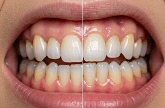
Often overlooked in their daily, tireless service, our teeth are marvels of natural engineering, each possessing a unique landscape of textures and surfaces. Far from being simple, uniform blocks, they present a fascinating array of contours, from glassy plains to intricate valleys and peaks. This diversity isn’t just for show; it’s intrinsically linked to their function, their development, and even how they interact with light to create a bright smile. Delving into the world of dental topography reveals a surprising level of detail and sophistication.
The Resilient Outer Layer: Enamel’s Smooth Facade
The part of the tooth we see most readily, the enamel, is the hardest substance in the human body, even tougher than bone. To the touch, healthy enamel often feels incredibly smooth, almost like polished porcelain. This smoothness is a key characteristic, contributing to its ability to resist the daily onslaught of chewing forces and to shed food debris. When you run your tongue over the front of your teeth, that sleek, almost glassy sensation is the enamel doing its job.
However, this apparent smoothness is a macroscopic observation. Under magnification, enamel reveals a more complex texture. One of the most fascinating micro-features are the perikymata. These are subtle, wave-like, horizontal grooves and ridges that mark the enamel surface. They are the external manifestations of the striae of Retzius, incremental growth lines formed during tooth development. On newly erupted teeth, perikymata can be more pronounced, giving the surface a slightly satiny or finely etched appearance rather than a perfectly flat polish. As we age, these features can gradually wear down, leading to a smoother, more uniform surface over time. The way light interacts with these microscopic features contributes significantly to the tooth’s natural luster and vitality.
Beneath the Sheen: Exploring Deeper Textures
Should the enamel be compromised or worn away, a different material, dentin, comes into play. Dentin forms the bulk of the tooth structure, lying directly beneath the enamel and also beneath the cementum that covers the roots. Compared to enamel, dentin is considerably softer and more porous. It’s composed of countless microscopic tubules, known as dentinal tubules, which radiate outward from the central pulp chamber. While not typically a visible or touchable surface in a completely intact tooth, its texture is inherently different – less dense and more yielding than the crystalline hardness of enamel. Its yellowish hue can also influence the overall color of the tooth if the overlying enamel is thin.
Covering the roots of our teeth is another specialized surface called cementum. This layer is softer than both enamel and dentin. Its texture is not designed for hardness or smoothness in the same way as enamel; instead, it’s slightly rougher. This characteristic is crucial because cementum serves as the attachment point for the periodontal ligaments, tiny fibers that anchor the tooth to the jawbone. This slightly irregular surface provides a better grip for these ligaments, ensuring the tooth remains securely in its socket. You wouldn’t typically feel cementum unless there’s gum recession, but its unique texture is vital for tooth stability.
The Working Surfaces: A Landscape of Function
The most complex and varied textures are found on the occlusal, or chewing, surfaces of our teeth, especially the molars and premolars. These surfaces are not flat; they are intricate landscapes designed for specific tasks like grinding, tearing, and crushing food.
Cusps: The Peaks of the Tooth
Cusps are the pointed or rounded elevations on the chewing surfaces. Think of them as the mountains or hills of the tooth. Molars typically have four or five prominent cusps, while premolars usually have two (hence their alternative name, bicuspids). Canines have a single, pointed cusp, perfect for their role in tearing. Incisors don’t have cusps in the same way but possess a sharp, incisal edge for cutting.
Grooves and Fissures: The Valleys and Crevices
Weaving between the cusps are grooves and fissures. These are depressions or developmental lines that create a network of valleys across the occlusal surface. Primary grooves, like the central groove on a molar, are major developmental lines that separate the main cusps. Smaller, supplemental grooves often branch off these, adding to the complexity. Fissures are often very narrow, deep clefts, particularly at the base of grooves. This intricate system helps to channel food away during chewing and provides escape pathways for food particles, making the grinding process more efficient.
Pits: Where Pathways Converge
At the intersection of developmental grooves, small, pinpoint depressions known as pits often form. The most common example is the central pit on a molar, typically found where multiple primary grooves meet. These pits can be quite deep and are areas where food debris and microorganisms can easily accumulate.
Ridges: The Elevated Pathways
Complementing the cusps and grooves are various types of ridges, which are linear elevations on the tooth surface.
- Marginal ridges are found on the mesial (towards the front of the mouth) and distal (towards the back of the mouth) borders of the occlusal surfaces of posterior teeth (molars and premolars). They also form the mesial and distal borders of the lingual (tongue-side) surface of anterior teeth (incisors and canines). These ridges act like rims, helping to contain food on the chewing surface.
- Triangular ridges descend from the tips of cusps towards the central area of the occlusal surface. They are named for their roughly triangular shape.
- When a buccal (cheek-side) triangular ridge and a lingual triangular ridge meet, they can form a transverse ridge, running across the occlusal surface.
- Some maxillary molars (upper molars) have an oblique ridge, which runs diagonally across the occlusal surface, a very distinctive feature.
The enamel surface, while appearing smooth to the naked eye, is covered in microscopic, wave-like ripples known as perikymata. These are the external manifestations of the striae of Retzius, which are incremental growth lines within the enamel itself. These subtle undulations play a crucial role in how light scatters across the tooth, contributing to its natural vibrancy and depth of appearance, especially in younger teeth.
Beyond the Chewing Surfaces: Other Textural Nuances
The non-chewing surfaces also have their own characteristic textures. The labial (lip-side) surfaces of anterior teeth and buccal (cheek-side) surfaces of posterior teeth are generally convex and relatively smooth. However, they may exhibit subtle developmental depressions or lobes, reflecting the way the tooth formed.
The lingual (tongue-side) surfaces are often more complex. Anterior teeth (incisors and canines) have a concave area called the lingual fossa, bordered by the aforementioned marginal ridges and a convex bulge near the gumline known as the cingulum. The texture here can feel quite different from the smooth outer surfaces, with defined edges and hollows. Lingual surfaces of posterior teeth are also generally convex but can feature grooves and other irregularities.
Proximal surfaces, those that face adjacent teeth (mesial and distal), are generally smoother to allow for tight contact points between teeth. These contact areas are crucial for maintaining the stability of the dental arch and preventing food from getting impacted between teeth. The texture here is less about active function and more about passive, stable interaction.
Developmental Signatures and Age-Related Transformations
Newly erupted permanent incisors often display a charming feature: mamelons. These are three small, rounded protuberances along the incisal edge. They are remnants of the developmental lobes from which the tooth formed. Mamelons typically wear away fairly quickly with normal use, leaving a straight, smooth incisal edge. Their presence is a tell-tale sign of a young, newly emerged tooth, contributing a distinct, scalloped texture to the biting edge.
Over a lifetime, the textures of our teeth inevitably change. The most common transformation is attrition, the gradual wearing down of tooth surfaces due to tooth-to-tooth contact during chewing or grinding. This can smooth out mamelons, reduce the height of cusps, and flatten the occlusal landscape. In some cases, wear can be significant enough to expose the underlying dentin, which, being softer, can wear at a different rate and present a different texture and color – often a duller, more matte finish compared to the gloss of enamel. The once prominent perikymata may become less distinct or disappear entirely on older, more worn enamel surfaces.
The journey across the surfaces of our teeth reveals an unexpected world of intricate design. From the broad, smooth sweeps of enamel to the complex mountain ranges of our molars, each texture and contour is a testament to the elegant functionality of the human body. These are not static structures but dynamic landscapes that develop, serve, and transform throughout our lives, each detail playing a part in the vital roles our teeth perform every single day.


