The human head, a marvel of biological engineering, is more than just a housing for our brain and the seat of our identity. It’s a complex assembly of bones, joints, and muscles working in intricate harmony. Central to many of its functions, from speaking to eating, is the dynamic relationship between the skull and the jaw. Understanding this anatomical partnership offers a fascinating glimpse into how our bodies are structured for everyday life.
The Protective Cranium
The skull itself can be broadly divided into two main parts: the cranium (or neurocranium) which encases the brain, and the facial skeleton (or viscerocranium) which forms the framework of our face. The cranium is not a single, solid bone but rather a collection of several flat and irregular bones fused together. These bones are the frontline defense for our most vital organ.
Key Cranial Bones
Several major bones form the protective vault of the cranium:
- Frontal Bone: This forms the forehead and the upper part of the eye sockets (orbits).
- Parietal Bones (Paired): These two large, flat bones form the majority of the roof and sides of the cranium.
- Temporal Bones (Paired): Located at the sides and base of the skull, inferior to the parietal bones. They house the structures of the ear and form a crucial part of the jaw joint.
- Occipital Bone: Situated at the back and base of the skull, it features a large opening called the foramen magnum, through which the spinal cord connects to the brain.
- Sphenoid Bone: Often described as butterfly-shaped, this complex bone sits at the base of the skull, anterior to the temporal and occipital bones. It articulates with almost every other cranial bone, acting as a keystone.
- Ethmoid Bone: A delicate, cuboidal bone located at the roof of the nose, between the two orbits. It contributes to the nasal cavity and the orbits.
These cranial bones are not seamlessly joined from birth. Instead, they are connected by fibrous joints called sutures. Prominent sutures include the coronal suture (between the frontal and parietal bones), the sagittal suture (between the two parietal bones), and the lambdoid suture (between the parietal bones and the occipital bone). In infancy, these sutures are more flexible and include larger membranous gaps called fontanelles (soft spots), allowing for brain growth and passage through the birth canal. They gradually ossify and fuse as we age.
The Framework of the Face – The Facial Skeleton
Anterior and inferior to the cranium lies the facial skeleton. These bones provide the structural support for our facial features, house our sensory organs like eyes and nose, and provide attachment points for facial muscles, enabling expressions. They also play a critical role in the initial stages of digestion and respiration.
Prominent Facial Bones
The facial skeleton includes several bones, but for understanding the jaw relationship, two are paramount:
- Maxillae (Paired, forming the Upper Jaw): These two bones fuse in the midline to form the upper jaw. They hold the upper teeth, form parts of the orbits, the nasal cavity, and the hard palate.
- Mandible (Lower Jaw): This is the largest and strongest bone of the face. It holds the lower teeth and is the only truly mobile bone of the skull (excluding the tiny ossicles of the middle ear). Its movement is central to chewing and speaking.
Other important facial bones include:
- Zygomatic Bones (Paired): Commonly known as the cheekbones, they form the prominence of the cheeks and contribute to the orbits.
- Nasal Bones (Paired): Small bones that form the bridge of the nose.
- Lacrimal Bones (Paired): Small, thin bones located in the medial wall of each orbit, housing the lacrimal sac (part of the tear drainage system).
- Palatine Bones (Paired): L-shaped bones located posterior to the maxillae, forming the posterior part of the hard palate and part of the nasal cavity and orbits.
- Inferior Nasal Conchae (Paired): Curved bones that project from the lateral walls of the nasal cavity, helping to warm and humidify air.
- Vomer: A single, thin bone that forms the posterior and inferior part of the nasal septum.
Spotlight on the Mandible – The Mover and Shaker
The mandible, or lower jawbone, is truly unique. It begins as two separate bones in fetal development (right and left hemi-mandibles) that fuse at the midline (symphysis menti) usually by the first year of life to form a single, U-shaped bone. Its mobility is what allows us to masticate (chew) food, speak, and make a wide range of facial expressions.
Key Features of the Mandible
The mandible has several distinct anatomical landmarks:
- Body: The horizontal, U-shaped portion that bears the lower teeth in sockets called alveolar processes. The chin, or mental protuberance, is the anterior prominence of the body.
- Ramus (plural: Rami): Two vertical projections, one on each side, extending upwards from the posterior part of the body.
- Angle of the Mandible: The junction where the posterior border of the ramus meets the inferior border of the body.
- Condylar Process: A posterior projection on the superior aspect of the ramus. It has a rounded head (the condyle) that articulates with the temporal bone to form the temporomandibular joint (TMJ).
- Coronoid Process: An anterior, triangular projection on the superior aspect of the ramus. It serves as an attachment site for the temporalis muscle, a major muscle of mastication.
- Mandibular Notch: The curved depression between the condylar and coronoid processes.
The Maxilla – The Stationary Anchor
While the mandible is known for its movement, the maxillae (plural of maxilla) form the stationary upper jaw. These paired bones are centrally located in the facial skeleton and articulate with many other facial and cranial bones, essentially anchoring the midface.
Notable Aspects of the Maxilla
Key features of the maxillae include:
- Alveolar Process: Similar to the mandible, this is the thickened ridge of bone that contains the tooth sockets (alveoli) for the upper teeth.
- Palatine Process: A horizontal plate that projects medially from each maxilla. These two processes meet in the midline to form the anterior three-quarters of the hard palate (the roof of the mouth and floor of the nasal cavity). The posterior part is formed by the palatine bones.
- Frontal Process: Extends superiorly to articulate with the frontal bone.
- Zygomatic Process: Projects laterally to articulate with the zygomatic bone.
- Maxillary Sinus: Each maxilla contains a large air-filled cavity, the largest of the paranasal sinuses.
The maxillae are critical not just for holding the upper teeth and forming part of the palate, but they also contribute significantly to the structure of the nasal cavity and the orbits of the eyes. Their central position and multiple articulations make them a keystone of the midfacial architecture. This intricate connection highlights the integrated nature of the skull’s design.
The Crucial Connection – The Temporomandibular Joint (TMJ)
The relationship between the skull and the jaw hinges, quite literally, on the Temporomandibular Joint (TMJ). This is a bilateral synovial joint, meaning there’s one on each side of the head, working in synchrony. It’s formed by the articulation of the mandibular condyle (from the mandible) with the mandibular fossa and articular tubercle of the temporal bone (part of the cranium).
The TMJ is one of the most complex joints in the body, capable of a remarkable range of movements. It’s not a simple hinge joint; it also allows for gliding and rotational movements. This complexity is facilitated by several structures:
- Articular Disc (Meniscus): A fibrocartilaginous disc located between the condyle and the temporal bone. This disc divides the joint cavity into two separate synovial compartments (upper and lower). It acts as a shock absorber, helps to stabilize the joint, and allows for smoother movements by conforming to the changing shapes of the articular surfaces during jaw motion.
- Joint Capsule: A fibrous capsule that encloses the joint, helping to retain synovial fluid, which lubricates the joint.
- Ligaments: Several ligaments, including the temporomandibular ligament, sphenomandibular ligament, and stylomandibular ligament, help to stabilize the joint, prevent excessive movement, and guide the condyle during function.
Movements of the Mandible via the TMJ
The TMJ facilitates several key movements of the mandible:
- Depression (Opening): Lowering the mandible. This involves both a hinge-like rotation in the lower joint compartment and an anterior gliding (translation) of the condyle and disc in the upper joint compartment.
- Elevation (Closing): Raising the mandible. This is the reverse of opening.
- Protrusion: Moving the mandible forward (anteriorly). This primarily involves gliding in the upper joint compartment.
- Retrusion (Retraction): Moving the mandible backward (posteriorly).
- Lateral Excursion (Side-to-Side): Grinding movements. This involves one condyle moving anteriorly and medially while the other condyle rotates.
The Power Behind the Movement – Muscles of Mastication
The sophisticated movements of the mandible are powered and controlled by a group of strong muscles known as the muscles of mastication. These muscles attach to both the cranium/facial skeleton and the mandible.
The Primary Masticatory Muscles
There are four primary pairs of muscles of mastication:
- Masseter Muscle: A powerful, thick, rectangular muscle located at the side of the face, overlying the ramus of the mandible. It originates from the zygomatic arch and inserts onto the lateral surface of the ramus and angle of the mandible. Its primary action is to elevate the mandible (close the jaw), and it also assists in protrusion.
- Temporalis Muscle: A large, fan-shaped muscle situated on the temporal fossa of the skull. It passes medial to the zygomatic arch and inserts onto the coronoid process and anterior border of the ramus of the mandible. It elevates the mandible and also retrudes it.
- Medial Pterygoid Muscle: Located deep to the ramus of the mandible, it runs roughly parallel to the masseter but on the medial side. It originates from the pterygoid plate of the sphenoid bone and inserts onto the medial surface of the ramus and angle of the mandible. It assists in elevating the mandible, protrusion, and side-to-side movements.
- Lateral Pterygoid Muscle: Situated superior to the medial pterygoid, this muscle has two heads. It runs horizontally and posteriorly from the sphenoid bone to the condylar process of the mandible and the articular disc of the TMJ. It is the primary muscle responsible for protruding the mandible and is crucial for opening the jaw (by pulling the condyle and disc forward). It also plays a key role in lateral excursions.
The coordinated action of these muscles is essential for effective chewing, speaking, and even yawning. Imbalances or issues with these muscles can significantly impact jaw function and comfort. Understanding their individual roles and synergistic actions is key to appreciating the biomechanics of the jaw and its overall health.
Dental Occlusion – Where Teeth Meet
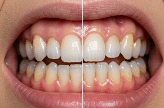
The relationship between the skull and jaw isn’t just about bones and joints; it culminates in how our teeth meet. Dental occlusion refers to the contact between the maxillary (upper) and mandibular (lower) teeth in any functional relationship. When we close our mouths, our teeth come together in a specific way, ideally allowing for efficient chewing and distributing forces evenly.
Proper occlusion is influenced by the skeletal relationship of the maxilla and mandible, the alignment of the teeth within each arch, and the guidance provided by the TMJs and muscles of mastication. The cusps and fossae (grooves) of opposing teeth are designed to interdigitate, much like gears, to grind and shear food. This intricate fit is vital not only for breaking down food but also for speech articulation and even the overall stability of the jaw system. Deviations from an ideal occlusion can sometimes be related to the underlying skeletal structure or the way the TMJ functions.
A Harmonious System
The anatomy of the human skull and its intricate relationship with the jaw is a testament to functional design. From the protective cranium housing our brain to the mobile mandible powered by robust muscles and guided by the complex TMJ, every component plays a vital role. The facial bones provide structure and support, while the precise interaction of the upper and lower jaws, culminating in dental occlusion, allows us to perform essential daily tasks like eating and speaking. Understanding these basic anatomical principles provides a foundation for appreciating the complexity and efficiency of this remarkable biological system. It’s a system where bone, joint, muscle, and teeth all work in concert, a finely tuned orchestra within our own heads.
