Content
Core Structures: The Building Blocks of the Root
Cementum: The Root’s Protective Outer Garment
Covering the entire outer surface of the root is a specialized, calcified tissue called cementum. Think of it as the root’s version of enamel, though it’s not nearly as hard. Its primary job is to provide a surface for the fibers of the periodontal ligament (which we’ll explore shortly) to attach, effectively anchoring the tooth into its bony socket. Cementum is continually, albeit slowly, deposited throughout life, a process that can help compensate for minor tooth wear or facilitate slight adjustments in tooth position. There are generally two main types: acellular cementum, which is formed first and covers roughly the cervical two-thirds of the root, and cellular cementum, typically found on the apical third and in areas where roots divide (furcations), which forms more rapidly and contains cells called cementocytes.Dentin: The Resilient Core
Beneath the thin layer of cementum lies dentin, which forms the substantial bulk of the root structure, just as it forms the bulk of the tooth’s crown underneath the enamel. Dentin is a hard, dense, yellowish-white tissue, more mineralized than bone but softer and more elastic than enamel. Its most characteristic feature is its microscopic structure, composed of countless tiny channels called dentinal tubules. These tubules radiate outwards from the central pulp cavity towards the cementum. Each tubule contains fluid and a long, slender cytoplasmic process from an odontoblast cell, whose body lines the pulp cavity. This tubular network is the reason why, if cementum is lost and dentin becomes exposed, teeth can become sensitive to temperature changes or certain foods.The Pulp Canal: Lifeline Through the Root
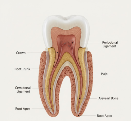
Running like a central corridor through the dentin in each root is the pulp canal, often referred to simply as the root canal. This narrow channel is a continuation of the pulp chamber, which is located in the crown of the tooth. The pulp canal extends from the floor of the pulp chamber down to the very tip, or apex, of the root. It’s not an empty space; rather, it’s precisely shaped to house the vital pulp tissue. The morphology of pulp canals can be surprisingly complex, varying in shape, size, and number even within a single tooth, presenting unique anatomical landscapes.Pulp Tissue: The Living Heart
Housed within the protective confines of the pulp chamber and the pulp canal(s) is the pulp tissue. This is the living, innermost part of the tooth, a soft, gelatinous connective tissue. It’s a rich environment, containing an intricate network of blood vessels (arterioles and venules), lymphatic vessels, nerves, and specialized cells, including odontoblasts (responsible for dentin formation) and fibroblasts. The blood vessels provide essential nutrients and oxygen to keep the tooth alive and responsive, while the nerves are primarily sensory, transmitting signals such as pain, temperature, and pressure, thereby acting as a biological alarm system for the tooth.Apical Foramen: The Gateway at the Tip
At the very end, or apex, of each root tip is a tiny, crucial opening called the apical foramen. This is the main portal through which the blood vessels, nerves, and lymphatic drainage from the surrounding jawbone enter and exit the pulp canal system. It’s a critical juncture, directly connecting the tooth’s internal, living environment with the rest of the body’s circulatory and nervous systems. While usually there’s one main foramen, it’s not uncommon to find multiple smaller openings, known as accessory foramina or an apical delta, especially in mature teeth, creating a more complex network at the root’s terminus.The Root’s Essential Support Network
The root itself is just one part of a sophisticated system designed to keep the tooth firmly in place and functioning correctly. Its immediate neighbors are just as important.Periodontal Ligament (PDL): The Tooth’s Suspension System
While not technically part of the tooth root itself, the periodontal ligament (PDL) is an absolutely indispensable component of the tooth root system. The PDL is a specialized, highly vascular and cellular connective tissue composed of countless tiny collagenous fibers. These fibers are precisely arranged to surround the root, effectively suspending the tooth in its bony socket. One end of these fibers (Sharpey’s fibers) embeds into the cementum of the root, and the other end embeds into the alveolar bone. This arrangement doesn’t just hold the tooth; it acts like a sophisticated shock absorber, cushioning the tooth and bone against the considerable forces of chewing and biting. The PDL also contains nerves that provide vital sensory information about tooth movement, pressure, and pain, helping to modulate bite force and protect the tooth from excessive loads. It also plays roles in tooth eruption and maintaining the physiological tooth position.The periodontal ligament is a truly remarkable tissue. It not only anchors the tooth securely but also allows for slight physiological movement, which is essential for distributing and dissipating chewing forces. Furthermore, it contains a rich supply of cells capable of forming new cementum and bone, contributing to ongoing repair and adaptation processes throughout an individual’s life, responding to functional demands.
Alveolar Bone: The Snug Socket
The alveolar bone, also known as the alveolar process, is the specialized part of the jawbone (either the maxilla in the upper jaw or the mandible in the lower jaw) that forms the bony sockets, or alveoli, which house the tooth roots. The integrity of this bone is crucial for tooth support. The PDL fibers embed directly into this bone on one side and into the root’s cementum on the other, creating a strong yet dynamic fibrous joint known as a gomphosis. The alveolar bone is constantly undergoing remodeling – breaking down and rebuilding – in response to the functional stresses placed upon it by the teeth. Its health and density are critical for the long-term stability and retention of the teeth.Not All Roots Are Created Equal: Embracing Variation
The fundamental components of the tooth root system are consistent across all teeth, but their specific configuration, number, and complexity can vary dramatically. This anatomical diversity is a key aspect of dental science and has practical implications for how teeth function and are cared for.Number of Roots: From Solo Acts to Multi-Rooted Ensembles
Teeth are often classified by the number of roots they possess, which generally correlates with their size and functional demands:- Single-rooted teeth: Typically, the anterior teeth (incisors and canines) and most premolars (bicuspids) in both arches have a single root. This simpler structure is well-suited for their primary functions of cutting, piercing, and tearing food.
- Multi-rooted teeth: Molars, designed for grinding and heavy mastication, almost always have multiple roots to provide a broader base for increased stability and resistance to occlusal forces. Upper molars commonly have three roots (two buccal, or cheek-side, and one palatal, or roof-of-the-mouth-side). Lower molars usually have two robust roots (one mesial, towards the front of the mouth, and one distal, towards the back). Some upper premolars, particularly the first premolars, can also be bi-rooted or even occasionally tri-rooted.