The living core of a tooth, known as the dental pulp, is a bustling hub of cellular activity, sensation, and defense. Like any active tissue in the body, it has an absolute requirement for a constant supply of nutrients and oxygen, and an equally efficient system for waste removal. This vital lifeline is provided by a surprisingly complex network of blood vessels that navigate the hard tissues of the tooth to reach its soft interior. Understanding this vascular system gives us a deeper appreciation for the tooth’s biology and its ability to respond to various stimuli.
The Journey Begins: Major Arterial Pathways
The story of how blood reaches the dental pulp starts much further up, with the major arteries of the head and neck. For all teeth, the initial source is the
common carotid artery, which branches in the neck. One of these branches, the
external carotid artery, is the primary highway for blood destined for the superficial structures of the head, including the jaws and, consequently, the teeth.
From the external carotid artery, a significant branch called the
maxillary artery takes over. This artery is a key player, responsible for supplying deep facial structures. It’s from the maxillary artery and its subsequent divisions that the specific vessels feeding individual teeth arise. The pathways diverge slightly depending on whether we are looking at the upper (maxillary) or lower (mandibular) teeth.
Supplying the Upper Teeth: Branches of the Maxillary Artery
For the maxillary teeth, several branches of the maxillary artery are involved. These are generally categorized as the superior alveolar arteries:
- Posterior Superior Alveolar Artery (PSAA): This artery typically arises from the maxillary artery before it enters the pterygopalatine fossa. It travels downwards and forwards, piercing the infratemporal surface of the maxilla to supply the molar and premolar teeth, as well as the surrounding gingiva and maxillary sinus lining. Multiple small branches from the PSAA enter the apical foramina (the tiny openings at the root tips) of these posterior teeth.
- Middle Superior Alveolar Artery (MSAA): The MSAA is not present in everyone, but when it is, it branches from the maxillary artery within the infraorbital canal or sometimes from the infraorbital artery itself. It descends within the lateral wall of the maxillary sinus to supply the premolar teeth and sometimes the mesiobuccal root of the first molar. It forms anastomoses (connections) with the posterior and anterior superior alveolar arteries, creating a network.
- Anterior Superior Alveolar Artery (ASAA): This artery is a branch of the infraorbital artery (which itself is a terminal branch of the maxillary artery). It courses downwards within the anterior wall of the maxillary sinus, giving off dental branches that supply the incisor and canine teeth. It also contributes to the blood supply of the nasal cavity and maxillary sinus.
These superior alveolar arteries form a complex plexus above the roots of the maxillary teeth, ensuring a rich and somewhat redundant blood supply to the pulp of each upper tooth.
Supplying the Lower Teeth: The Inferior Alveolar Artery
For the mandibular teeth, the pathway is a bit more straightforward after the maxillary artery. A major branch, the
inferior alveolar artery (IAA), descends to enter the mandibular foramen on the medial surface of the ramus of the mandible. Before entering this foramen, it often gives off a mylohyoid branch.
Once inside the mandible, the inferior alveolar artery travels through the mandibular canal, running beneath the roots of the mandibular teeth. Along its course, it gives off numerous small
dental branches. These dental branches ascend towards the root apices of the molars, premolars, canines, and incisors of the lower jaw, entering the pulp through their apical foramina. The main trunk of the inferior alveolar artery continues forward and terminates by dividing into the mental artery (which exits through the mental foramen to supply the chin and lower lip) and the incisive artery (which continues within the bone to supply the anterior teeth and anastomoses with its counterpart from the other side).
The dental pulp’s vitality is critically dependent on its specialized vascular network. This system diligently delivers essential oxygen and nutrients while simultaneously removing metabolic waste products. Without this continuous and carefully regulated exchange, the delicate pulp tissue cannot survive or function, underscoring the fundamental role of these blood vessels in maintaining tooth health.
Entering the Inner Sanctum: Passage into the Pulp Chamber
The primary route for these nutrient-carrying arteries to enter the tooth’s pulp chamber is through the
apical foramen, a small opening (or sometimes multiple small openings) located at the very tip (apex) of each tooth root. An arteriole, typically one or two main ones depending on the root, passes through this foramen alongside nerves and venules.
In addition to the apical foramen,
accessory canals can also provide pathways for blood vessels. These are smaller channels that can branch off the main root canal and exit along the side of the root. While less significant in terms of total blood volume compared to the apical foramen, they represent alternative or supplementary routes for vascular supply and can be clinically relevant.
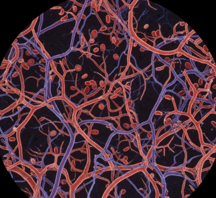
The Microcirculation: Lifeblood of the Pulp
Once inside the pulp chamber and root canals, the entering arterioles begin to branch extensively, forming a sophisticated microcirculatory network. This is where the real work of nutrient and gas exchange happens.
The arterioles subdivide into smaller vessels called
terminal arterioles, which then give rise to a dense
capillary network. This network is particularly rich in the coronal pulp (the part of the pulp in the crown of the tooth) and especially concentrated in a zone just beneath the odontoblasts, the cells responsible for forming dentin. This subodontoblastic capillary plexus is crucial for supporting the high metabolic activity of the odontoblasts as they maintain and repair dentin.
Pulp capillaries are typically fenestrated, meaning they have small pores or windows in their walls, which facilitates the efficient exchange of nutrients, oxygen, and waste products between the blood and the surrounding pulp tissue. The structure of this capillary bed is not static; it can adapt to changing physiological needs and respond to stimuli.
In some areas, arteriovenous anastomoses (AVAs) or shunts may exist. These are direct connections between arterioles and venules that can bypass the capillary bed, potentially playing a role in regulating blood flow and pressure within the confined space of the pulp chamber. However, the exact extent and functional significance of AVAs in human dental pulp are still areas of ongoing research.
After passing through the capillary beds, the blood, now depleted of oxygen and rich in carbon dioxide and metabolic wastes, is collected into
venules. These venules gradually merge to form larger veins that will carry the blood away from the tooth.
Venous Drainage: The Return Journey
The venous drainage system of the dental pulp largely mirrors the arterial supply. Small venules within the pulp coalesce to form larger veins that exit the tooth primarily through the apical foramen, accompanying the arteries and nerves.
For the
mandibular teeth, the dental veins drain into the
inferior alveolar vein, which travels alongside the inferior alveolar artery within the mandibular canal. This vein then typically drains into the
pterygoid venous plexus, a network of veins located in the infratemporal fossa, near the pterygoid muscles. The pterygoid plexus has multiple connections, including to the maxillary vein and facial vein.
For the
maxillary teeth, venous drainage is also directed towards the pterygoid venous plexus via veins that correspond to the superior alveolar arteries (posterior, middle, and anterior superior alveolar veins). Some drainage may also occur via the infraorbital vein and connect with the facial vein.
Ultimately, blood from both the maxillary and mandibular regions, after passing through networks like the pterygoid plexus, will find its way into larger veins such as the
maxillary vein, the
facial vein, and then into the
retromandibular vein, which contributes to the internal or external jugular veins, returning the blood towards the heart.
Regulation of Pulpal Blood Flow
The blood flow to the dental pulp is not a passive process; it is actively regulated to meet the tissue’s metabolic demands and respond to various stimuli. This regulation is achieved through a combination of neural and local factors.
Nervous control primarily involves sympathetic adrenergic nerve fibers that accompany the blood vessels. Activation of these sympathetic nerves generally leads to vasoconstriction (narrowing of the blood vessels), which can reduce blood flow. There is also evidence for parasympathetic and sensory nerve involvement in modulating pulpal blood flow, creating a complex interplay.
Local factors, such as changes in oxygen levels, carbon dioxide levels, pH, and the presence of inflammatory mediators, can also significantly influence vessel diameter and blood flow. For instance, an increase in metabolic activity or the initial stages of inflammation might lead to vasodilation (widening of blood vessels) to increase the supply of nutrients and defensive cells.
This intricate regulatory system is vital because the pulp is housed within a rigid, unyielding chamber of dentin. Unlike soft tissues elsewhere in the body that can swell when blood flow increases significantly (as in inflammation), the pulp has very limited space to expand. Therefore, precise control of blood flow is essential to prevent excessive increases in intrapulpal pressure, which could compress blood vessels and compromise the pulp’s own blood supply—a dangerous feedback loop.
In essence, the journey of nutrients to the dental pulp is a marvel of anatomical engineering. From large arteries in the neck to microscopic capillaries woven throughout the tooth’s core, this vascular system ensures that the cells responsible for a tooth’s vitality, sensation, and defense are continually nourished and sustained. The complexity of this supply underscores the biological dynamism of what might otherwise seem like a simple, inert structure.