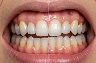
The gleaming, hard outer layer of our teeth, known as enamel, stands as the body’s most mineralized and durable tissue. Its primary role is to protect the sensitive inner parts of the tooth from the daily onslaught of chewing forces, temperature changes, and acid attacks from bacteria. While the vast majority of enamel is composed of inorganic hydroxyapatite crystals, a fascinating and crucial, albeit small, percentage is made up of organic material, predominantly proteins. These proteins are not mere bystanders; they are the unsung architects and regulators during enamel’s complex formation process, a dance known as amelogenesis.
Understanding these protein components offers profound insights into how this remarkable biological ceramic is constructed, why it sometimes forms imperfectly, and even how we might one day repair or regenerate it. Though largely removed once enamel matures, their transient presence dictates the final structure and integrity of this vital protective shield.
Before diving into the specific proteins, it’s essential to appreciate the context of amelogenesis. This intricate process is carried out by specialized cells called ameloblasts. These cells secrete a protein-rich extracellular matrix, which then guides the nucleation and growth of hydroxyapatite crystals. As the enamel layer thickens and matures, most of these proteins are systematically degraded and reabsorbed by the ameloblasts, leaving behind the densely packed mineral structure we recognize as mature enamel.
The Dominant Family: Amelogenins
By far the most abundant proteins in the developing enamel matrix are the
amelogenins, constituting about 80-90% of the total protein content during the secretory stage. Encoded by the AMELX gene (located on the X chromosome, with a homologous copy, AMELY, on the Y chromosome in many mammals), these proteins are somewhat enigmatic but absolutely critical. Amelogenins are hydrophobic and prone to self-assembly, forming spherical nanostructures or “nanospheres.”
The prevailing theory is that these amelogenin nanospheres act as spacers and organizers, controlling the size, shape, and orientation of the growing hydroxyapatite crystals. They prevent premature fusion of individual crystals, ensuring the development of long, slender, highly organized crystallites that are characteristic of enamel’s unique prismatic architecture. Think of them as temporary scaffolding that dictates the blueprint of the final mineralized structure. As mineralization progresses, amelogenins are gradually cleaved by proteinases and removed, allowing the crystals to grow larger and pack more tightly together, leading to the extreme hardness of mature enamel.
Mutations in the AMELX gene are a primary cause of X-linked amelogenesis imperfecta, a hereditary condition characterized by thin, soft, or discolored enamel, underscoring the indispensable role of amelogenins in forming healthy enamel.
Mature human tooth enamel is remarkably acellular, being the most mineralized tissue in the body. It is composed of approximately 96% inorganic mineral by weight, primarily hydroxyapatite. The remaining percentage consists of organic material, mostly proteins, and water, which are crucial during its development.
The Supporting Cast: Key Non-Amelogenin Proteins
While amelogenins take center stage in terms of sheer volume, several other non-amelogenin proteins play equally vital, albeit more specialized, roles in the enamel formation symphony. These proteins often work in concert with amelogenins and each other, contributing to different facets of matrix organization and mineralization.
Ameloblastin: The Adhesion Expert
Ameloblastin (AMBN), also known as amelin or sheathelin, is the second most abundant protein in the developing enamel matrix, though it makes up a much smaller fraction compared to amelogenins (around 5-10%). Encoded by the AMBN gene, ameloblastin is thought to play a crucial role in maintaining the integrity of the enamel layer and ensuring proper adhesion of ameloblasts to the forming enamel surface. It seems to be particularly important for controlling the organized structure of enamel rods (or prisms) and the interrod enamel that surrounds them.
Studies have shown that ameloblastin helps to define the rod boundaries and is involved in cell signaling, influencing ameloblast differentiation and function. Its absence or malfunction leads to severe enamel defects, often characterized by enamel separating from the underlying dentin or a complete failure of enamel formation, highlighting its structural and signaling importance. It is rapidly processed and degraded, meaning its presence is more transient but no less important.
Enamelin: The Crystal Nucleator
Enamelin (ENAM) is another critical non-amelogenin protein, encoded by the ENAM gene. Though present in smaller quantities than ameloblastin, it is believed to be one of the first proteins secreted by ameloblasts at the very beginning of enamel formation, near the dentin-enamel junction (DEJ). Its primary proposed function is in initiating the nucleation of hydroxyapatite crystals and promoting their elongation.
Enamelin is thought to bind tightly to the mineral crystals, possibly serving as a template or scaffold upon which the initial mineral deposition occurs. Its early expression and localization at the DEJ suggest it is fundamental for laying down the foundational layer of enamel. Mutations in the ENAM gene can cause various forms of amelogenesis imperfecta, ranging from thin, hypoplastic enamel to enamel that is poorly mineralized, demonstrating its vital role in both the quantity and quality of enamel produced.
Tuftelin: The Early Bird
Tuftelin (TUFT1) is expressed early during tooth development, even before amelogenin and ameloblastin, primarily at the dentin-enamel junction. Its precise role has been a subject of debate, but it’s hypothesized to be involved in the initial mineralization events at the interface between enamel and dentin. It might also play a role in ameloblast differentiation and signaling.
While tuftelin is rapidly degraded, some fragments persist in mature enamel, particularly in structures called enamel tufts, which are hypomineralized, ribbon-like features that project from the DEJ into the enamel. These tufts are relatively rich in protein, and tuftelin is a significant component. The persistence of tuftelin in these structures suggests it might have a long-term, albeit minor, structural or modulatory role in mature enamel, or it could simply be a remnant of early developmental processes.
The Clean-Up Crew: Proteinases in Enamel Maturation
The transformation of the soft, protein-rich enamel matrix into hard, mineralized tissue requires the precise and timely degradation and removal of the scaffold proteins. Two key enzymes, or proteinases, are responsible for this crucial “clean-up” phase:
MMP-20, also known as enamelysin, is secreted by ameloblasts throughout the secretory stage of amelogenesis. Its primary job is to perform the initial, specific cleavage of enamel matrix proteins, particularly amelogenin, ameloblastin, and enamelin. These initial cleavages are thought to be important for processing the proteins into forms that can properly assemble and function within the matrix, and also to prepare them for later, more extensive degradation. MMP-20 essentially “pre-digests” the matrix, preventing it from becoming overly aggregated or dysfunctional before the final maturation hardening begins.
Kallikrein-4 (KLK4)
As enamel formation transitions from the secretory to the maturation stage, the expression of MMP-20 decreases, and another proteinase,
Kallikrein-4 (KLK4), takes over. KLK4 is a serine proteinase with a much broader digestive capability. It is responsible for the bulk degradation of the already-processed enamel matrix proteins, clearing them out to make space for the hydroxyapatite crystals to grow and interlock. The efficient removal of this proteinaceous material by KLK4 is absolutely essential for achieving the high degree of mineralization that gives mature enamel its exceptional hardness. Failure of KLK4 activity leads to hypomineralized enamel that is soft and prone to wear, as seen in some forms of amelogenesis imperfecta.
Disruptions in the production, processing, or removal of any of these critical enamel proteins during tooth development can lead to structural enamel defects. These conditions, collectively known as amelogenesis imperfecta, can manifest as enamel that is abnormally thin, soft, rough, pitted, or discolored. Such defects significantly compromise the tooth’s protective function.
Proteins in Mature Enamel: More Than Just Remnants?
While the vast majority of organic material is removed during maturation, mature enamel is not entirely devoid of proteins. Trace amounts, constituting less than 1% by weight, remain. These are often found concentrated in specific microstructural features like enamel tufts, lamellae (crack-like features), and at the sheaths surrounding enamel rods. The protein profile of mature enamel is different from that of developing enamel, consisting mainly of more insoluble proteins and peptides, likely degradation products or tightly bound components.
Historically, these residual proteins were often considered mere remnants trapped within the mineral. However, ongoing research suggests they might play subtle roles in modulating enamel’s mechanical properties, such as its fracture toughness, or influencing its interaction with the oral environment, including demineralization and remineralization processes. The exact nature and function of these persistent proteins in mature enamel are still areas of active investigation, but their presence indicates that the story of enamel proteins doesn’t entirely end with amelogenesis.
The Broader Significance
The study of enamel proteins is far from just an academic exercise. It holds the key to understanding the etiology of inherited enamel defects like amelogenesis imperfecta, paving the way for better diagnosis and potentially future gene-based therapies. Furthermore, by dissecting nature’s own strategies for building this incredibly resilient biomaterial, scientists are gaining inspiration for developing novel biomimetic approaches to dental repair and regeneration. Imagine being able to regrow enamel or create restorative materials that perfectly mimic its structure and properties – understanding enamel proteins is a fundamental step towards such futuristic goals.
In conclusion, the protein components of tooth enamel, though minor in the mature tissue, are the masterminds behind its formation. From the voluminous amelogenins shaping crystal growth to the precise actions of ameloblastin, enamelin, and the enzymatic duo of MMP-20 and KLK4, each protein plays an orchestrated and indispensable role. Their intricate interplay ensures the development of enamel’s unique structure, providing a lifetime of protection for our teeth. The ongoing exploration of these molecular architects continues to unveil the secrets of this natural marvel.


