Content
Delving Deeper: The Dentin Layer
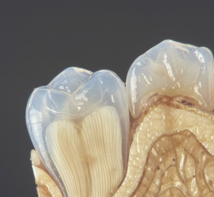
Beneath the formidable shield of enamel lies dentin, a layer that constitutes the main bulk of the tooth. If enamel is the armor, dentin is the supportive structure underneath. It extends from the crown, where it is covered by enamel, down into the root, where it is typically covered by another material called cementum. While also a hard, calcified tissue, dentin is considerably softer than enamel. Its composition is different too; it is about 70 percent inorganic material, mainly hydroxyapatite, 20 percent organic material, largely collagen fibers, and 10 percent water. This higher organic content, particularly the collagen, gives dentin a degree of resilience that enamel lacks, helping to prevent fractures of the overlying brittle enamel. A hallmark of dentin is its microscopic structure, characterized by countless tiny channels known as dentinal tubules. These tubules radiate outwards from the central pulp cavity towards the enamel or cementum. Within these tubules reside extensions of specialized cells called odontoblasts, whose cell bodies line the pulp cavity. This makes dentin a living, sensitive tissue. It is through these tubules that sensations like hot, cold, or touch can be transmitted to the nerve endings in the pulp, sometimes resulting in tooth sensitivity. Dentin is typically pale yellow, and its shade significantly influences the overall color of the tooth, as it is visible through the translucent enamel. Unlike enamel, dentin can be formed throughout life. The initial dentin is called primary dentin. Secondary dentin is formed slowly after tooth eruption, gradually reducing the size of the pulp chamber. Tertiary dentin, also known as reparative dentin, can form in response to stimuli like wear or irritation, providing a localized protective barrier.The Root’s Covering: Understanding Cementum
Moving down from the crown and into the area below the gum line, we encounter cementum. This specialized calcified substance forms a thin layer covering the entire root surface of the tooth, much like enamel covers the crown. Its primary role is quite distinct from enamel or even dentin. Cementum serves as the crucial attachment medium for the periodontal ligament fibers, which are tiny strands of tissue that anchor the tooth securely within its bony socket in the jaw. Think of it as the binding agent that holds the tooth in place. In terms of hardness, cementum is softer than both dentin and enamel, more comparable to bone in its physical properties and composition. It consists of about 45 to 50 percent inorganic material (hydroxyapatite) and 50 to 55 percent organic matter (mainly collagen) and water. There are two main types of cementum: acellular and cellular. Acellular cementum is the first to be formed and covers the cervical third or half of the root, closer to the crown. It forms before the tooth erupts and does not contain cells within its matrix. Cellular cementum, on the other hand, is formed after tooth eruption and is found predominantly on the apical half of the root and in areas between roots of multi-rooted teeth. It contains cells called cementocytes, which are entrapped cementoblasts (cementum-forming cells), residing in spaces called lacunae, similar to osteocytes in bone. Cementum is avascular, meaning it has no direct blood supply, receiving its nutrients from the surrounding periodontal ligament. A fascinating aspect of cementum is its ability to undergo continuous deposition throughout life. This allows for some degree of repair and adaptation to tooth movement or wear, helping to maintain the integrity of the tooth’s attachment.Cementum, Enamel, Dentin: A Tale of Three Tissues
While enamel, dentin, and cementum all contribute to the structure and function of a tooth, they possess distinct characteristics that set them apart. Understanding these differences helps appreciate the intricate design of our dental anatomy.Hardness and Mineral Content
The most striking difference lies in their hardness, directly related to their mineral content. Enamel stands out as the undisputed champion of hardness, boasting approximately 96 percent mineral content, primarily hydroxyapatite. This makes it exceptionally resistant to wear. Dentin, with around 70 percent mineral content, is significantly softer than enamel but harder than cementum. Its greater proportion of organic material, like collagen, imparts some flexibility. Cementum is the softest of the three, with a mineral content of about 45 to 50 percent, making it comparable to bone. This relative softness means it can wear away more easily if exposed due to gum recession.Location and Coverage
Their placement within the tooth is also a key differentiator. Enamel exclusively covers the anatomical crown of the tooth, the portion normally visible. Dentin forms the core structure, underlying the enamel in the crown and the cementum in the root. It constitutes the largest part of the tooth. Cementum is specifically confined to the root surfaces, extending from the cementoenamel junction, where enamel and cementum meet near the gum line, to the apex or tip of the root.Cellularity and Vitality
The presence or absence of living cells greatly influences their nature. Enamel is acellular; it contains no living cells and is therefore considered a non-vital tissue. It cannot repair itself. Dentin is a vital tissue because it contains the cytoplasmic extensions of odontoblasts, cells whose bodies line the pulp cavity. These processes within the dentinal tubules make dentin sensitive. Cementum can be either acellular, primarily near the crown-root junction, or cellular, more apically and in interradicular areas, containing cementocytes. These cells, similar to bone cells, help maintain the cementum layer and can contribute to its repair and continued deposition.Regenerative and Reparative Capabilities
Their ability to regenerate or repair varies significantly. Enamel has no inherent ability to regenerate once it is fully formed and mineralized. Lost enamel is lost permanently unless restored through other means. Dentin, being a vital tissue, has some reparative capacity. Odontoblasts can lay down secondary dentin throughout life and tertiary, or reparative, dentin in response to injury or irritation. Cementum also demonstrates a dynamic nature. It is continuously deposited throughout life, particularly cellular cementum. This allows for the adaptation of the tooth’s attachment apparatus and some repair of root surface damage.Primary Functions
Each layer serves a primary, specialized function:- Enamel: Its main job is to provide a hard, durable surface for mastication (chewing) and to protect the underlying dentin from physical, thermal, and chemical assaults.
- Dentin: It provides foundational support for the enamel, prevents enamel fracture due to its slight elasticity, protects the pulp, and transmits stimuli from the tooth surface to the pulp. It also largely determines tooth color.
- Cementum: Its principal function is to provide attachment for the periodontal ligament fibers, thus anchoring the tooth in its socket. It also plays a role in root repair and helps compensate for occlusal wear by continued apical deposition.
Color and Permeability
Enamel is translucent, its color ranging from light yellow to grayish white, influenced by its thickness and the color of the underlying dentin. It is semipermeable, allowing certain ions and small molecules to pass through. Dentin is typically a light to dark yellow and is more opaque than enamel. Its permeability is due to the dentinal tubules, which can act as pathways for fluids and other substances if the protective enamel or cementum is breached. Cementum is light yellow and slightly opaquer than dentin. It is also permeable, especially the cellular type.It is quite remarkable how these three distinct tissues work in concert. Enamel provides the tough exterior shield, dentin offers a resilient core and pathway for sensory feedback, while cementum ensures the tooth remains firmly anchored within the jaw. Each layer is uniquely structured and composed for its specific role in the overall function of a tooth.In essence, the tooth is a complex structure, far more intricate than a simple piece of bone. The subtle yet significant differences and collaborative functions of enamel, dentin, and cementum allow our teeth to withstand incredible forces, respond to various stimuli, and remain anchored throughout life. Recognizing their unique properties helps us appreciate the sophisticated biological engineering at play within each tooth and the importance of maintaining the integrity of these layers.